ORAL AND MAXILLOFACIAL SURGERY
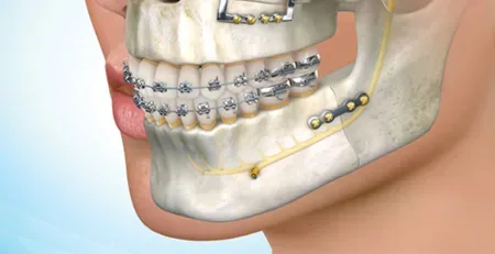
Oral and maxillofacial surgery is the specialty field that performs various surgical procedures on the teeth and surrounding tissues. Some of the commonly performed procedures are as follows:
• TOOTH EXTRACTION
The procedure most commonly performed by oral and maxillofacial surgeons is tooth extraction in adults and children. Teeth may need to be extracted due to decay, impaction, periodontal disease, infection, etc.
Can’t any dentist extract my tooth?
All dentists receive the basic training required for tooth extraction. However, advances in modern dentistry and preventive dentistry practices have made it possible for far fewer teeth to be extracted today than in the past. Although most dentists still perform tooth extractions, they refer their patients to oral and maxillofacial surgery specialists for better surgical outcomes. The experience of oral and maxillofacial surgeons enables them to perform extractions more quickly and with less trauma to the surrounding tissues.
• APICAL RESECTION
The term apical resection (root amputation) means cutting and removing the tip of the root and eliminating the inflammatory tissue or cyst at the root apex. It is generally performed for inflammatory formations at the root tips of anterior teeth, in cases where root canal treatment alone cannot be successful. It is the process of cleaning the root tip of the tooth and the inflammatory/cystic tissue surrounding it down to the healthy bone tissue around it, and filling and closing the root canal.
• EXPOSURE OF AN IMPACTED TOOTH FOR ERUPTION
The canine teeth in the upper jaw normally erupt between the ages of 11 and 13. Because these teeth erupt later, in some cases they may not fit into the dental arch and remain impacted within the jawbone. They are often located on the inside of the palate, behind the front teeth. Exposing an impacted tooth is usually a simple procedure. The gum and, if necessary, bone over the impacted tooth are removed to expose the tooth. Then an orthodontic bracket is bonded to the tooth, and with the help of a chain attached to this bracket, the orthodontist brings the tooth into the correct position.
• ODONTOGENIC (TOOTH-ORIGIN) INFECTIONS
Infections originating from the tooth are called odontogenic infections. Following loss of vitality of the tooth, bacteria cause an infection around the root apex. The infection may follow the path of least resistance within the bone, pass into the soft tissue, and spread farther from the source of the tooth. The oral and maxillofacial surgeon’s duty, with a command of head and neck anatomy, is to accurately predict the routes of spread of these infections and, when necessary, bring the infection under control by ensuring drainage of the abscess.
• JAW CYSTS
Cysts are pathological cavities located within the jawbones or surrounding soft tissues, surrounded by a wall and filled with liquid or semi-liquid materials. Cysts in the jaws are often tooth-related. These cysts, which usually form around the tooth root, can reach large sizes if left untreated. Since medications are not effective in the treatment of cysts, they must be removed surgically as a whole (with their sac).
• REMOVAL OF AN IMPACTED TOOTH
By the age of 18, the average adult has a total of 32 teeth, 16 in the lower jaw and 16 in the upper jaw. Each tooth has a specific name and function. The teeth at the front of the mouth (incisors, canines, and premolars) function to bite and tear food. The back teeth, namely the molars, are used to grind food and prepare it for swallowing. However, there is room for only 28 teeth in an average mouth. Therefore, the eruption of the remaining teeth can be painful for patients. These teeth, located furthest back among the molars, are called third molars or wisdom teeth.
Why should wisdom teeth be extracted?
Wisdom teeth are the last teeth to erupt in the mouth. If there is enough space in the jaw for these teeth and they can take their place in the dental arch with healthy gum tissue, there is no need to extract them. Unfortunately, this is a rare situation. Commonly, wisdom teeth cannot erupt properly and need to be extracted. As impacted teeth search for a suitable path to erupt, they may take different positions within the bone. They may erupt sideways, emerge partially through the gum, or remain completely impacted beneath the gum and bone.
Teeth that cannot erupt properly create various problems. In cases of partial impaction, the gum around the tooth provides a favorable environment for bacteria to multiply, and as a result, infection develops. The infection then manifests in the patient as swelling and pain. It is also a common belief that the pressure from an erupting wisdom tooth can disrupt the alignment of the teeth in front of it. The most serious problem is the formation of cysts or tumors around impacted teeth; as a result, healthy bone tissue and teeth are damaged. Extraction of the impacted tooth in question is generally sufficient to resolve these problems. Early extraction of impacted teeth that are expected not to erupt is preferred in order to prevent these issues.
• DENTAL IMPLANT
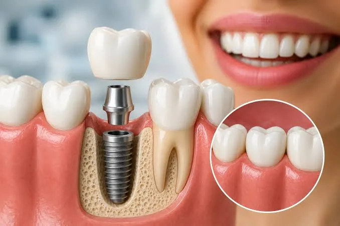
Dental implants are changing people’s lifestyles. Designed to form the foundation for porcelain restorations that look and function like natural teeth, this product gives a person who has lost a natural tooth the opportunity to eat and smile as they wish.
What is a dental implant?
Implants are artificial tooth roots made of titanium that are surgically placed into the jawbone in the area where tooth loss has occurred. Titanium integrates with the bone (fuses with it), creating a solid foundation for the tooth to be placed on top. Then, reduced tooth-shaped parts are screwed into the implant and extend out of the gum, and the tooth loss in the area is restored together with the porcelain tooth to be placed on top. At the same time, implants help preserve the patient’s facial structure by preventing bone loss caused by the jawbone remaining without teeth.
Surgical Procedure
Placement of implants is possible through a one- or two-stage surgical procedure. First, the implant is placed into the jawbone under local anesthesia. Following this surgical procedure, the implants are left beneath the gum for 2 to 4 months to allow them to integrate with the jawbone. During this period, if possible, the patient is fitted with a temporary prosthesis and advised to consume soft foods.
When the implant integrates with the jawbone, the second stage begins. Under local anesthesia, a small incision is made in the gum over the implant to access it, and a part extending out of the gum is screwed into the implant. This part, called a “gingival former,” prevents the gum from growing back over the implant while also allowing the gum to take on a natural shape around it, like the tissue around a natural tooth. In this way, the porcelain tooth to be placed on the implant looks natural, as if it were emerging from the gum. About one week after the gingival former is placed, impressions are taken from the implant and sent to the laboratory. In the following sessions, various try-ins are performed, such as checking the fit of the framework and determining the shape and color of the tooth. When the porcelain tooth reaches its final form, the gingival former is removed from the implant and a reduced tooth-shaped abutment is screwed into the implant. The prepared tooth is bonded to this abutment, completing the treatment.
What types of prostheses can be made on implants?
To restore a single missing tooth, a single crown prosthesis (tooth crown) is made on the implant. In cases of multiple missing teeth, enough implants can be placed in the edentulous area and a fixed bridge-type prosthesis can be made on them. The number of implants to be placed is determined by the dentist, taking into account factors such as the number of missing teeth, the condition of the jawbone, and what type of prosthesis will be made. For example, if there are two adjacent missing teeth, 2 implants are required; if there are 3 adjacent missing teeth, 2 to 3 implants are required. When all teeth in a jaw are missing, if a bridge-type fixed prosthesis is planned, 6 to 8 implants are needed, whereas if an implant-supported removable prosthesis is planned for the same jaw, 2 to 4 implants are sufficient. Removable prostheses attach to the implants through prepared sockets inside them, while bridge prostheses are completely fixed and can only be removed by the dentist.
Why should I prefer dental implants over traditional prostheses?
There are several reasons for this: First of all, there is no need to grind the adjacent teeth to fill the gap left by the missing tooth. While the tooth placed on the implant offers you the closest experience to your natural tooth, the implant itself functions as a root within the bone, helping prevent bone resorption over the years and preserve its volume. In addition, by providing retention for removable prostheses, it eliminates the difficulty of using especially lower dentures and improves the patient’s quality of life.
So, am I a candidate for implant treatment?
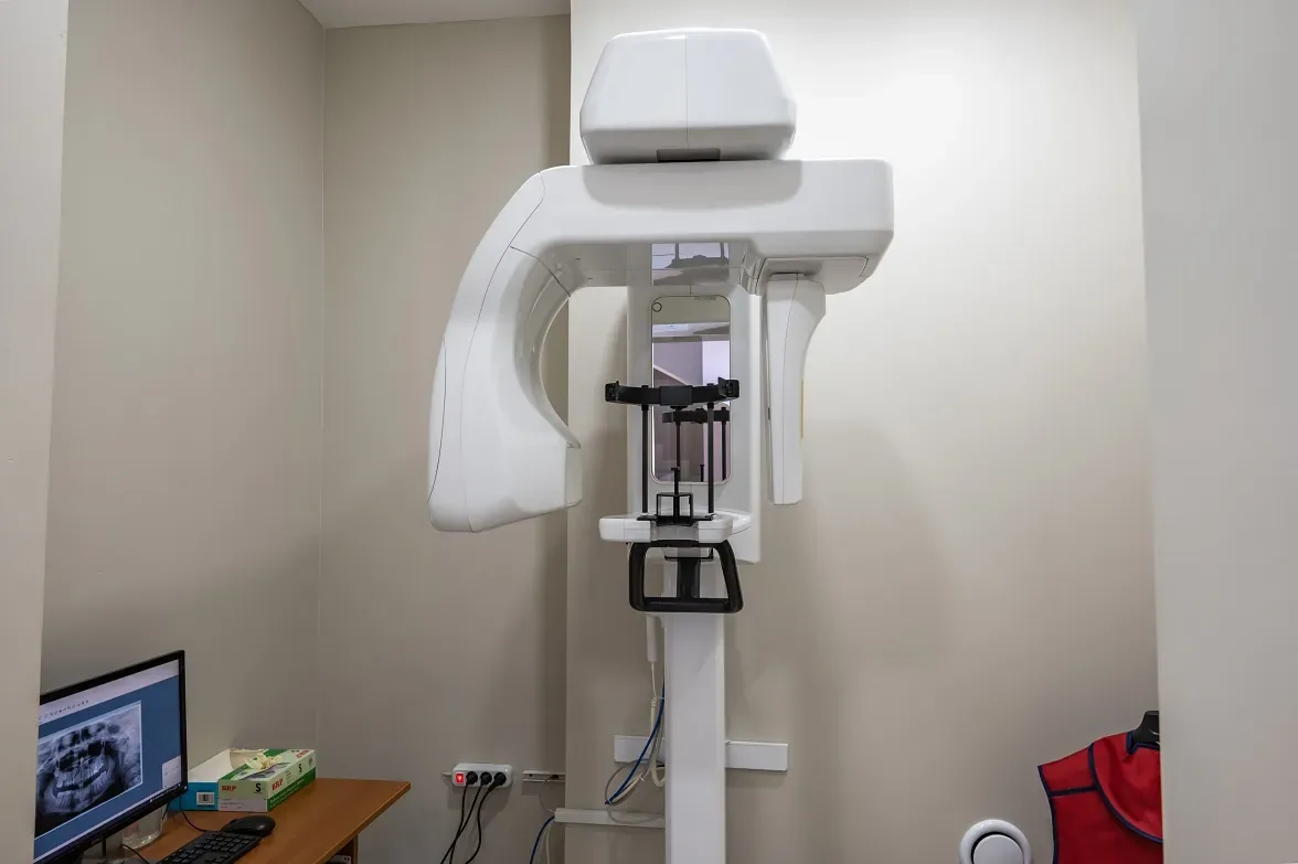
If you want to have implants, your medical history should be evaluated in addition to a comprehensive intraoral and radiological examination. If your mouth is not suitable for implant treatment, treatments that can make it suitable (such as bone grafting, etc.) are recommended to you. Before treatment begins, all risks, benefits, and treatment options are evaluated, and the most appropriate treatment plan is determined for each patient.
Do implants require special care?
If well cared for after placement, implants will serve you for many years. For this reason, it is necessary to take time to maintain good oral hygiene (brushing and flossing) and to visit the dentist at regular intervals.
• BONE AUGMENTATION
As time passes after tooth loss, volume loss occurs in the bone in the affected area. In cases where tooth loss has been prolonged, the bone may resorb to the point where implants cannot be placed.
Today, in areas with insufficient bone, it is often possible to increase the volume of the bone. In this way, implants of appropriate length and diameter can be placed, providing functional and aesthetic restoration.
While small bone defects or extraction sockets can be filled with artificial bone powders called bone grafts, in cases of severe bone loss it may be necessary to perform bone augmentation by taking bone from another area of the jaw or from the hip.
Sinus lift procedure
The maxillary sinuses are air-filled spaces located inside the cheeks and above the upper molars. The roots of some upper teeth can extend into the sinus. When such teeth are extracted, a thin layer of bone remains between the inside of the mouth and the sinus. Since dental implants are supported by bone tissue, there must be sufficient bone thickness in the area where they are placed. If the sinus floor is too thin, implant placement is not possible.
In this case, it becomes possible to place implants by performing a sinus lift (sinus lifting) procedure. In this procedure, the sinus membrane is lifted by entering through the lateral wall of the sinus in the area where the teeth were extracted, and bone powder is placed between the sinus floor and the sinus membrane. Depending on the bone height, implants can also be placed in the same session; or the implant placement procedure is performed 4 months later.
In the period when the sinus lift option was not available, patients with drooping sinuses had to use removable prostheses, whereas thanks to sinus lift, most patients have gained the opportunity to use a bridge in the upper back region.
• PRE-PROSTHETIC SURGERY
Preparation of the mouth before prosthesis fabrication is called “pre-prosthetic surgery.” Some patients may need minor surgical procedures before full or partial denture fabrication in order to provide a high level of comfort. Since the prosthesis rests on the bone tissue, it is important that the bone has the proper shape and size. For example, after extraction, bone spurs may remain in the relevant area. In such cases, these spurs must be removed so that the prosthesis fits well.
The procedures commonly needed to prepare the mouth for a prosthesis are as follows: • Smoothing bone spurs • Bone augmentation • Trimming excess gum tissue • Deepening the gums Each patient is evaluated individually, and the necessary procedures are determined according to the diagnosis and expectations.

Doka Klinik Dental Team
This article is for information only; please consult our dentists for a treatment plan tailored to you.